THE LAB
Micro X-Ray Computed Tomography (MXCT) is a non-destructive technique that visualizes inner features in the opaque solid objects. The 3-D geometries are computer re-constructed from the series of 2-D X-ray pictures acquired during the stepwise rotation of the investigated object. They represent internal density variations in the sample.
The technique can scrutinize wide range of materials, e.g.:
- rocks
- ceramics
- metals
- integrated circuits
- bio or medical samples
High-resolution, micro X-ray CT differs from conventional medical CAT-scanning in its ability to resolve details as small as few microns in size, even when imaged objects are made of high density and low-contrast materials.
The facility utilizes a newly purchased X-Radia 400 instrument. The X-Ray source (on the left side) is a 150 keV/10 W.
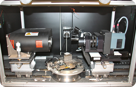
The instrument is capable of accepting nominally large samples (up to 0.5 m in length; though only of fairly low density). The technique is ideal for oil and gas drilling feasibility studies. The system supports larger and heavier core samples for pore connectivity and microstructure modeling.
To streamline the data acquisition and processing a second workstation was added to the setup. It is loaded with analysis software (reconstruction, viewing, post-analysis statistics, etc.). Both Image-J and Blob-3D programs are available for data analysis.
User Manual: The updated user manual is accessible here: : (pdf)
Special Procedures: The descriptions of additional procedures related to measurement, calibration, processing, and analysis are accessible here: (pdf)